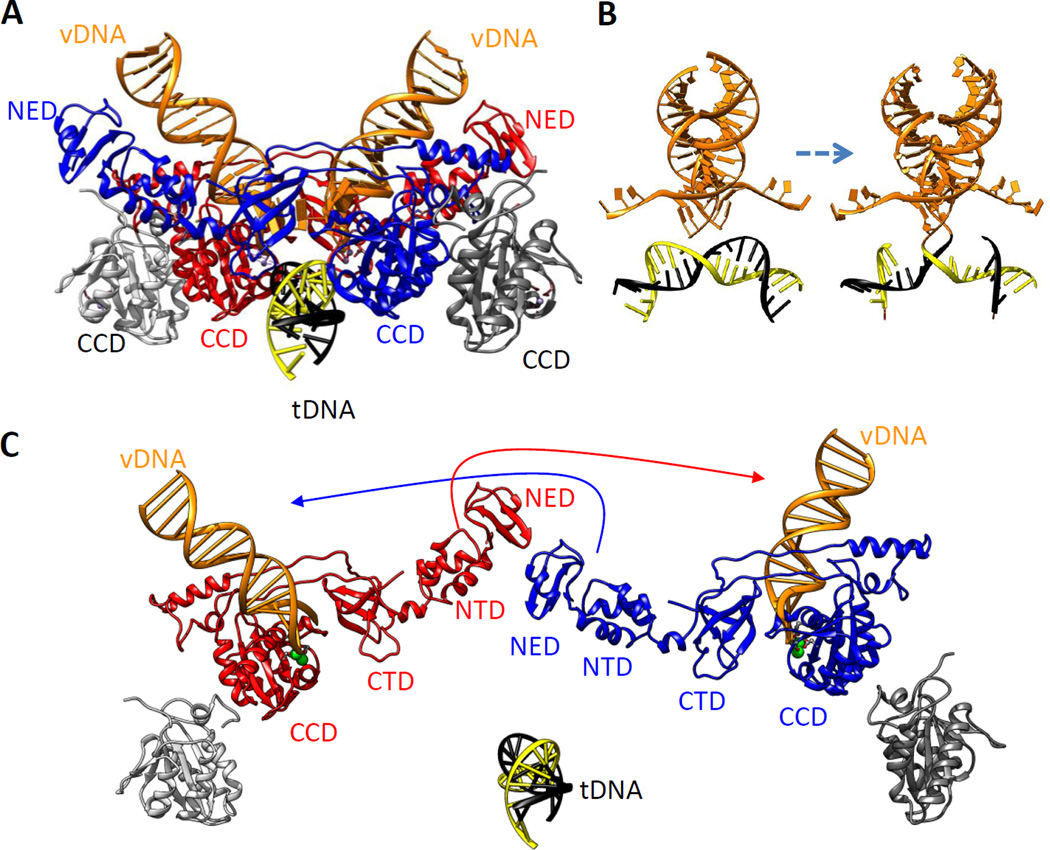
Figure 3.
PFV IN tetramer in complex with two vDNA oligonucleotides and a tDNA. (a) The assembled complex (PDB 4E7K) (89; see also supplemental movies in 88) is shown in ribbon representation with the inner subunits in red and blue. Only the CCDs of the outer subunits (gray) are resolved. vDNA oligonucleotides are in orange ribbon ladder representation and the tDNA in yellow and black. The locations of the NEDs and CCDs are indicated. (b) DNA components of the complex portrayed before and after joining, rotated 90° about the y axis from panel a. Processed vDNA ends are shown prior to joining (left) and after joining (right) to the target DNA. (c) The complex shown in panel a is pulled apart to show the positions of all domains in the inner subunits. Interactions between the distal NTD and NED of one inner subunit and the vDNA held in the CCD of the other inner subunit are indicated by arrows. Assembly of the complex is shown in Video 1. Abbreviations: CCD, catalytic core domain; CTD, C-terminal domain; IN, integrase; NED, N-terminal extension domain; NTD, N-terminal domain; PFV, prototype foamy virus; tDNA, target DNA; vDNA, viral DNA.

