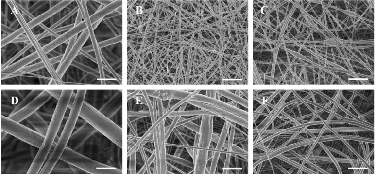
Figure 2.
SEM images showing the morphology of fibers mat electrospun from gelatin/PCL solution (A), gelatin/PCL solution containing 10 wt % nHA (B), and gelatin/PCL solution containing 10 wt % bone powder (C). Images are shown at different magnifications to reveal surface structures ((D), (E) and (F) correspond to (A), (B) and (C), respectively). Scale bar: (A), (B) and (C), 10 μm; (D), (E) and (F), 2 μm.

