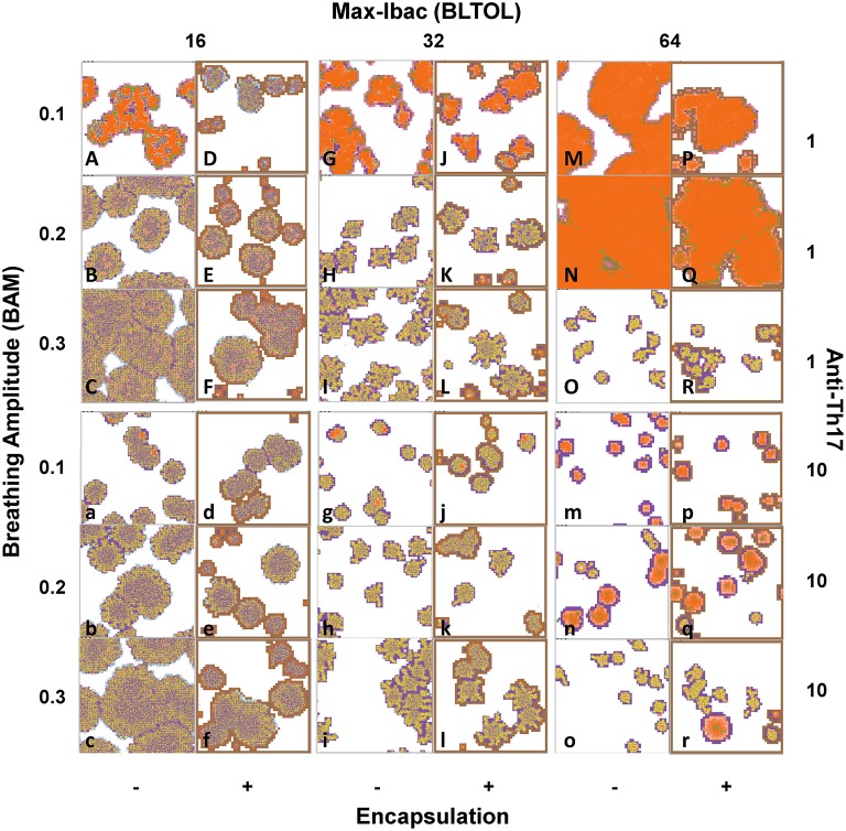
Figure 10.
Phenotypical characteristics of the lesions after running the program until day 46 post-challenge. Type A.- Proliferative in progression, with low quantities of Dbac and a high proportion of Kbac (pictures B, C, F, b, c and f). Type C.- Exudative, based on a large necrotic center surrounded by an active ring of alveoli filled with PMNs and the massive presence of Dbac (pictures A, G, J, M, N, P and Q). Type D.- Exudative controlled, with smaller lesions than in type C surrounded by a ring of aAMs (pictures g, j, m, p, n, q and r). Type B.- Proliferative controlled, as there is a very low quantity of Dbac and a very high proportion of Kbac (all other pictures). Colors: Alveoli: White: when a single AM is present; Black: when the AM is destroyed; Blue: when the AM is destroyed and replaced by another AM from the interstitium; Violet: when an activated AM (aAM) is present; Pink: when neutrophils occupy the alveoli; Brown: capsule. Bacilli: Blue challenge bacilli; Red: Intracellular bacilli (Ibac); Green: Extracellular (Ebac); Brown: growing Ebac; Orange: dormant (Dbac); Yellow: killed bacilli (Kbac).

