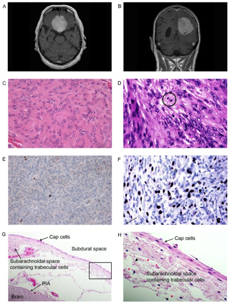
Figure 1.
Tumors of the two patients used for sequence analysis. Tumor from patient 1 (IA) presented to the left (A, C and E), and tumor from patient 2 (II) to the right (B, D and F); (A) and (B) show the Magnetic resonance imaging (MRI) for patients 1 and 2 (Table 1); (C) Meningothelial meningioma grade I with lobules of uniform meningioma cells with typical intranuclear inclusions. Hematoxylin and eosin stain (HE), 400×; (D) Atypical meningioma grade II with atypical features and scattered mitoses. HE, 600×; (E) Very few immunoreactive tumor cells for the proliferation marker Ki-67 in this grade I tumor, 200×; (F) Elevated activity for the proliferation marker Ki-67 in this grade II meningioma (200×); (G) The normal arachnoid contains an external layer of horizontally oriented cap cells (200×). Traversing the subarachnoid, a loose web-like tissue containing trabecular cells, fibrous tissue and vessels that fuse with the inner Pia mater covering the brain surface; (H) The enlarged marked section in G at 400× showing the horizontally oriented external cap cells in the arachnoid membrane.

