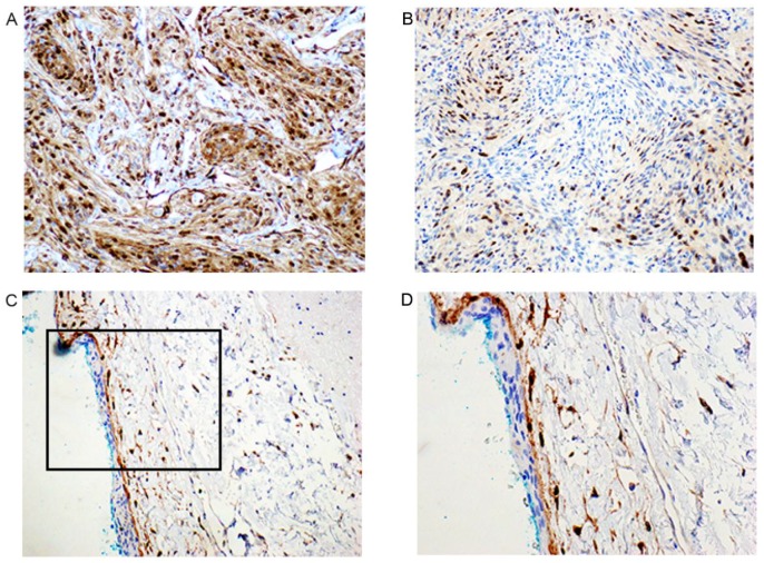
Figure 4.
Immunohistochemistry of Cyclin D1 (A–D) in meningioma (A–B) compared to normal arachnoid membranes (C–D). (A) Meningioma with strong nuclear signal of Cyclin D1 expression (200×); (B) Another tumor with moderate signal for the nuclear Cyclin D1 expression (200×); (C) Immunohistochemistry Cyclin D1 in arachnoid membrane from cadaver with no meningioma. The external cap cell layer of the arachnoid membrane is negative, while some of the trabecular cells in the internal layer stain positive (200×); (D) The same as shown in C with higher magnification (400×).

