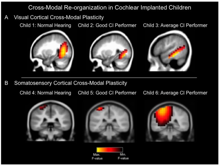
Figure 1.
(A) Current density reconstructions for the cortical visual evoked potential (CVEP) P2 component in three different children: a child with normal hearing (Child 1), a cochlear implanted (CI) child who is a good performer with good speech perception (Child 2), and a cochlear implanted child who is an average performer with an average speech perception score (Child 3). The visual stimulus consisted of a visual motion stimulus. Yellow regions demonstrate the localization of cortical sources with higher statistical likelihood, whereas darker regions reflect cortical sources with lower statistical likelihood. The normal hearing child (Child 1) and good CI user (Child 2) show significant sources only in expected higher-order visual processing regions in response to the visual motion stimulus, whereas the average CI user (Child 3) demonstrates evidence of cross-modal plasticity where the visual motion stimulus results in significant cortical sources in higher-order visual as well as temporal (auditory) cortical areas; (B) Current density reconstructions for the cortical somatosensory evoked potential (CSSEP) N70 component in 3 children: A child with normal hearing (Child 4), a cochlear implanted child with a good speech perception score (Child 5), and a cochlear implanted child with an average speech perception score (Child 6). Vibrotactile stimulation of the right index finger results in significant cortical sources only in somatosensory cortex in the normal hearing child (Child 4) and good CI user (Child 5), whereas the average CI user (Child 6) demonstrates significant sources in somatosensory cortex, as well as temporal (auditory) cortical regions, indicative of cross-modal re-organization [23].

