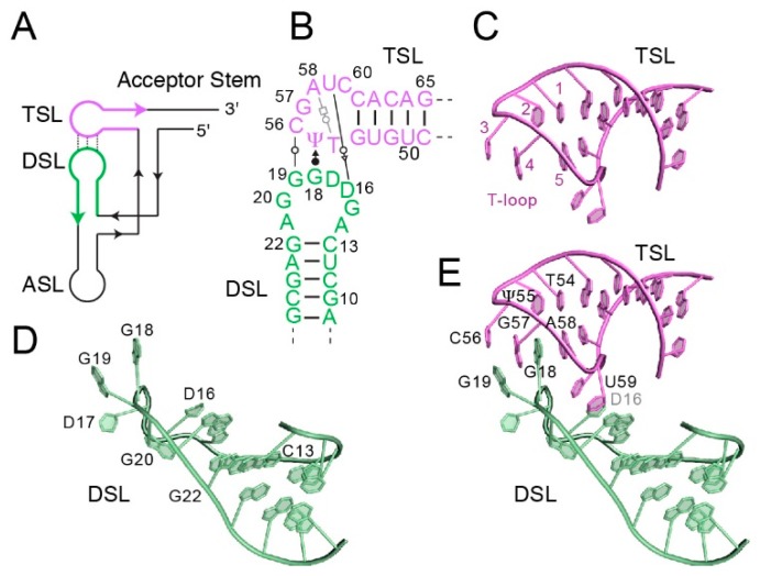
Figure 1.
Structure of the tRNA elbow. (A) Schematic of the connectivity of tRNA. ASL, anticodon stem-loop. DSL, D-stem-loop. TSL, T-stem-loop. (B) Secondary structure of the yeast tRNAPhe elbow. Non-canonical pairs between the D- and T-loops are depicted with Leontis-Westhof [29] symbols. Residue numbering reflects the tRNA convention. (C) Structure of the T-loop of yeast tRNAPhe (PDB ID 1EHZ). Note the unstacked residue 56 corresponding to position 3 of the generalized T-loop, and the gap between residues 57 and 58, corresponding to positions 4 and 5 of the generalized T-loop. (D) Structure of the D-loop of yeast tRNAPhe. Dihydrouridines are located at residues 16 and 17. (E) Interaction of D- and T-loops forms the elbow. Note inter-loop base pairs between residues 19 and 56, and intercalation of D-loop residue 18 into the T-loop. Structure figures were prepared with PyMol [30].

