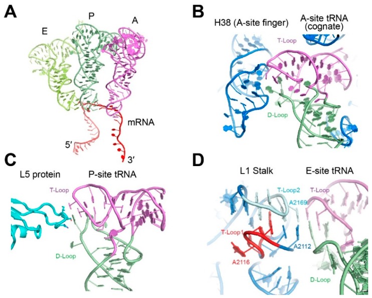
Figure 2.
Interaction of the tRNA elbow with the ribosome. (A) Relative positions of the three classical state tRNAs from ribosome cocrystal structures. (PDB ID 4V6F). (B) Interaction of the tRNA elbow with helix 38 (the “A-site finger”). (PDB ID 4V6F) rRNA is in blue. (C) Interaction of the tRNA elbow with the L5 protein (cyan) in the P-site. (PDB ID 4V51). (D) Interaction of the tRNA elbow with the L1 stalk in the E-site. (PDB ID 4V4I). The two interdigitated T-loops of the L1 stalk are denoted T-Loop1 and T-Loop2 in the 5′ to 3′ direction.

