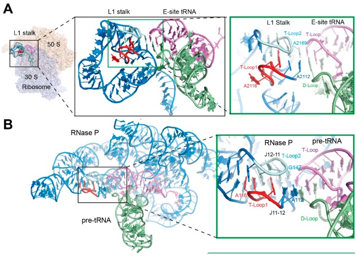
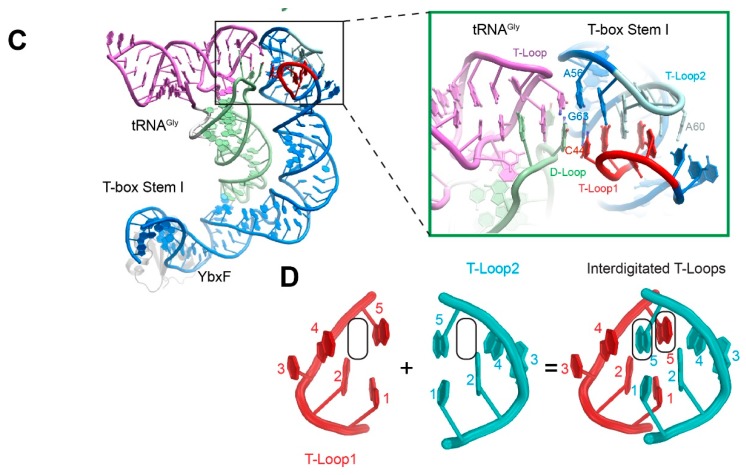
Figure 3.
Recognition of the tRNA elbow by the interdigitated T-loop motif. (A) The 5′ and 3′ interdigitated pentaloops of the ribosomal L1 stalk are colored red and cyan, respectively. The tRNA D- and T-loops are pale green and violet, respectively. (PDB ID 4V4I). (B) Structure of RNase P holoenzyme bound to pre-tRNA. (PDB ID 3Q1Q). (C) Structure of a glycine-specific T-box Stem I domain bound to its cognate tRNAGly. (PDB ID 4LCK). (D) Interdigitation of two head-to-tail pentanucleotide T-loops (red and cyan, respectively) forms a densely packed core structure. The five nucleotides that form each T-loop are numbered and the stacking gaps denoted by the rounded rectangles.


