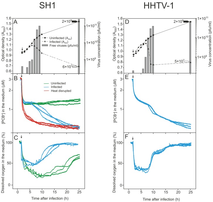
Figure 2.
Growth parameters and physiological changes in Haloarcula hispanica during infection by (A–C) SH1 and (D,F) HHTV-1 (MOI of 20). Unadsorbed viruses were removed at 2 h p.i. and measurements were begun at 2 h 15 min p.i. (indicated by arrows) and were carried out in MGM medium at 37 °C with aeration. (A,D) Turbidities of the infected and uninfected cultures; the number of free progeny viruses (pfu/mL) in the infected cultures; and the number of viable cells (cfu/mL) at 25 h p.i. in the uninfected (black arrow head) and infected cultures (white arrow head); (B,E) PCB− binding in the presence of PCB− (calibrated with 3 µM PCB−) to infected, uninfected, and heat disrupted (showing the maximal binding) Har. hispanica cells (n = 3); (C,F) The level of dissolved oxygen in the medium of infected and uninfected Har. hispanica cells (n = 3).

