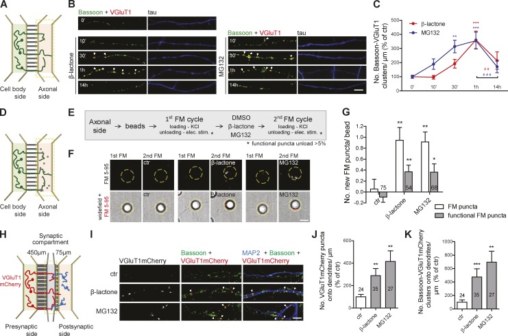
Figure 1.
Axonal proteasome inhibition boosts formation of presynaptic sites. (A) Microfluidic devices. (B) Local proteasome inhibition by 10 µM β-lactone and 1 µM MG132 (box in A) induced an increase in presynaptic clusters (arrowheads), assessed by immunostaining for the SV marker VGluT1 (red) and the active zone marker Bassoon (green). (C) Number of presynaptic clusters (colocalized VGluT1 and Bassoon punctum >0.05 µm2) per axonal length (percentage of 0 min). Three to five independent experiments. (D and E) Formation of new functional presynaptic sites on beads by repeated cycles of FM dye staining. (F) New FM dye puncta (red) appear on beads (dashed circles) after local β-lactone or MG132 treatment (1 h; box in D). (G) Number of new total and active FM puncta per bead. n, beads. (H) Synapse formation chambers. (I) Proteasome inhibition in the synaptic compartment (box in H) triggered presynaptic assembly (arrowheads) onto dendrites, assessed by presynaptic expression of VGluT1mCherry and staining for MAP2 (blue) and Bassoon (green). (J and K) Number of VGluT1mCherry puncta (J) and presynaptic clusters (K) along dendrites per length of transduced axons (percentage of control). n, dendrites. (G, J, and K) Three independent experiments. ***, P < 0.001; **, P < 0.01; and *, P < 0.05 compared with 0 min or control and ###, P < 0.001 and ##, P < 0.01 (Kruskal-Wallis test followed by Dunn’s multiple comparison test). Results are presented as mean values ± SEM. Bars, 5 µm.

