Figure 1.
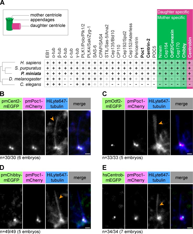
Identification and characterization of centriole markers in starfish. (A) Overview of identified starfish (P. miniata) homologues of centriolar and PCM proteins compared with those of select model organisms. Black plus sign indicates the presence of a clear homologue; gray plus sign indicates functional homology with divergent amino acid sequence. Mother centriole–specific markers are shown in green and daughter centriole–specific markers are shown in pink (see also schematic representation on the top left). Genes coding for proteins shown in bold were used in this study. (B–E) Fluorescent protein fusions of indicated centriolar proteins were validated as live cell markers in the ciliated epithelium of starfish embryos. HiLyte647 tubulin labels the cilium (orange arrowheads). pmPoc1-mCherry and mEGFP-pmCentrin-2 label both mother and daughter centrioles; pmOdf2-mEGFP and pmChibby-mEGFP are mother-specific markers; hsCentrobin-mEGFP is a daughter-specific marker. Maximum intensity projections of 2–5 confocal sections are shown. Bars, 1 µm.

