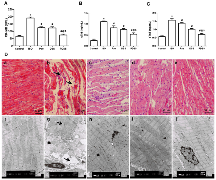
Figure 2.
Effect of PDSS on ISO-induced changes in serum-specific cardiac injury biomarkers [(A) CK-MB, (B) cTnI, (C) cTnT; values are expressed as mean ± S.D. (n = 8). *P < 0.001 vs. Control; #P < 0.001 vs. ISO; @P < 0.001 vs. Pae; $P < 0.001 vs. DSS (one-way ANOVA).] and on ISO-induced histopathologic changes of heart [(D) (a) control, (b) ISO, (c) Pae, (d) DSS and (e) PDSS; (a–e) belongs to light microscopic study; heart tissues were stained with hematoxylin and eosin and visualized under light microscope at ×200 magnification. (D) (f) control, (g) ISO, (h) Pae, (i) DSS and (j) PDSS; (f–j) belongs to TEM study; heart tissues were stained in alcohol uranyl acetate and lead citrate and viewed under transmission electron microscope at ×10000 magnification].

