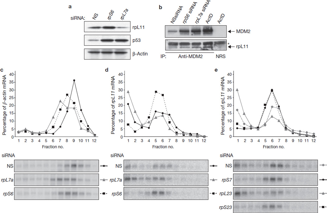
Figure 4.
Depletion of 40S ribosomal proteins results in translational upregulation of the rpL11 mRNA. (a) Western blots showing the levels of rpL11, p53 and β-actin proteins in cells transfected for 48 h with NSsiRNA, rpS6 siRNA2 or rpL7a siRNA. (b) Western blots showing the levels of rpL11 and MDM2 present in immunoprecipitates (IP) by using either an anti-MDM2 antibody or normal rabbit serum (NRS). Extracts used in the immunoprecipitation are from cells transfected for 24 h with NSsiRNA or with rpS6 or rpL7a siRNAs, or treated with actinomycin D (ActD) at 10 ng ml−1 for 10 h. The asterisk indicates the light chain of the anti-MDM2 antibody, which reacts with the secondary antibody. (c, d) Northern blot analysis of β-actin mRNA (c) and rpL11 mRNA (d) distribution on polysomes of A549 cells transfected with the indicated siRNAs for 30 h. (e) Northern blot analysis of rpL11 mRNA distribution on polysomes of A549 cells transfected with the indicated siRNAs. Fractions 4–12 in c–e contained translating polysomes. Uncropped images of blots are shown in Supplementary Information, Fig. S5.

