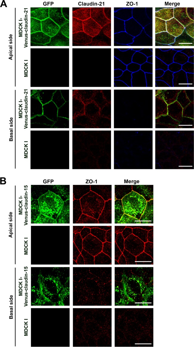
FIG 1.
Mouse claudin-21 localization in MDCK I transfectant clones. (A) MDCK I cells or Venus-claudin-21-expressing transfected MDCK I cells (MDCK I-Venus-claudin-21 cells) were cultured to confluence on glass coverslips and examined by confocal laser scanning microscopy. The cells were triple stained with an anti-GFP pAb, an anti-claudin-21 pAb, and an anti-ZO-1 MAb. The anti-GFP-positive signals overlapped the anti-claudin-21-positive signals and the anti-ZO-1-positive signals. Stacked images of the apical or basal side of the epithelial cells are shown. Green, GFP; red, claudin-21; blue, ZO-1. Bars, 10 μm. (B) MDCK I cells or Venus-claudin-15-expressing transfected MDCK I cells (MDCK I-Venus-claudin-15 cells) were cultured to confluence on glass coverslips and examined by confocal laser scanning microscopy. The cells were stained with an anti-GFP pAb and an anti-ZO-1 MAb. The anti-GFP-positive signals overlapped the anti-ZO-1-positive signals. Stacked images of the apical or basal side of the epithelial cells are shown. Green, GFP; red, ZO-1. Bars, 10 μm.

