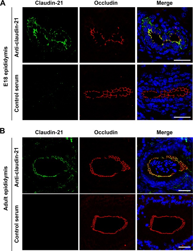
FIG 4.
Claudin-21 expression in mouse tissues. Immunofluorescence micrographs show epididymides from an E18 mouse (A) and an adult mouse (B) costained with an anti-claudin-21 pAb and an antioccludin MAb or with antiserum and an antioccludin MAb. DAPI (4′,6-diamidino-2-phenylindole) was used to detect nuclei. The anti-claudin-21- or antiserum-positive signals (green), the antioccludin-positive signals (red), and nuclei (blue) are shown. Bars, 50 μm.

