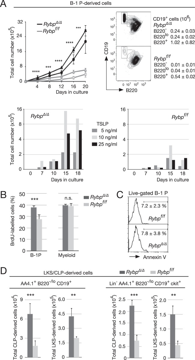
FIG 4.
Increased TSLP-activated self-renewal and expansion of isolated, HSC- or CLP-derived Rybp mutant B-1 progenitors. (A) Expansion and phenotype of cultured B-1 progenitors (B-1P; Lin− B220−/lo CD19+ ckit+). (Top left) Proliferation kinetics of 2 × 10³ B-1P sorted from bone marrow of Rybpf/f MxCre+ or Rybpf/f MxCre− mice previously injected with poly(I·C) (circles) or of Rybpf/f; Cre-ERT2 mice after treatment with 4′-OHT (RybpΔ/Δ) or vehicle (ethanol; Rybpf/f) during the first 24 h in culture (triangles). Cells were cocultured with S17 stromal cells seeded in Transwells, with no cell contact, and medium supplemented with 10 ng/ml TSLP. (Top right) Representative flow cytometry plots of CD19- and B220-labeled cells after 20 days in culture; data correspond to mean absolute numbers and SD of CD19+ cells with different B220 labeling (boxes depicted in plots; triplicate cultures of three independent experiments). (Bottom) A total of 2 × 10³ B-1P sorted from pooled RybpΔ/Δ (left) and Rybpf/f (right) bone marrow (n = 3 mice/genotype per experiment) were cultured in the presence of the indicated amounts of TSLP. Cells were collected and counted at the indicated time points (means of duplicate cultures from two independent experiments). (B) BrdU-labeled B-1P and myeloid progenitors (MyP) isolated from mice of the indicated genotypes 12 h after intraperitoneal injection of 1 mg BrdU. (C) Apoptosis within the B-1P population of the indicated genotypes, assessed by FITC-conjugated annexin V labeling of propidium iodide-impermeable cells. Horizontal bars denote apoptotic cells showing mean and SD values from triplicates of two independent experiments. (D) Enhanced production of B-1P from LKS and CLP pools in mice of the indicated genotypes. (Left) Total B220−/lo cells; (right) B-1P. Bar graphs show mean values and SD from triplicates of two independent experiments. **, P ≤ 0.01; ***, P ≤ 0.001; ****, P ≤ 0.0001); n.s., not statistically significant (P ≥ 0.05).

