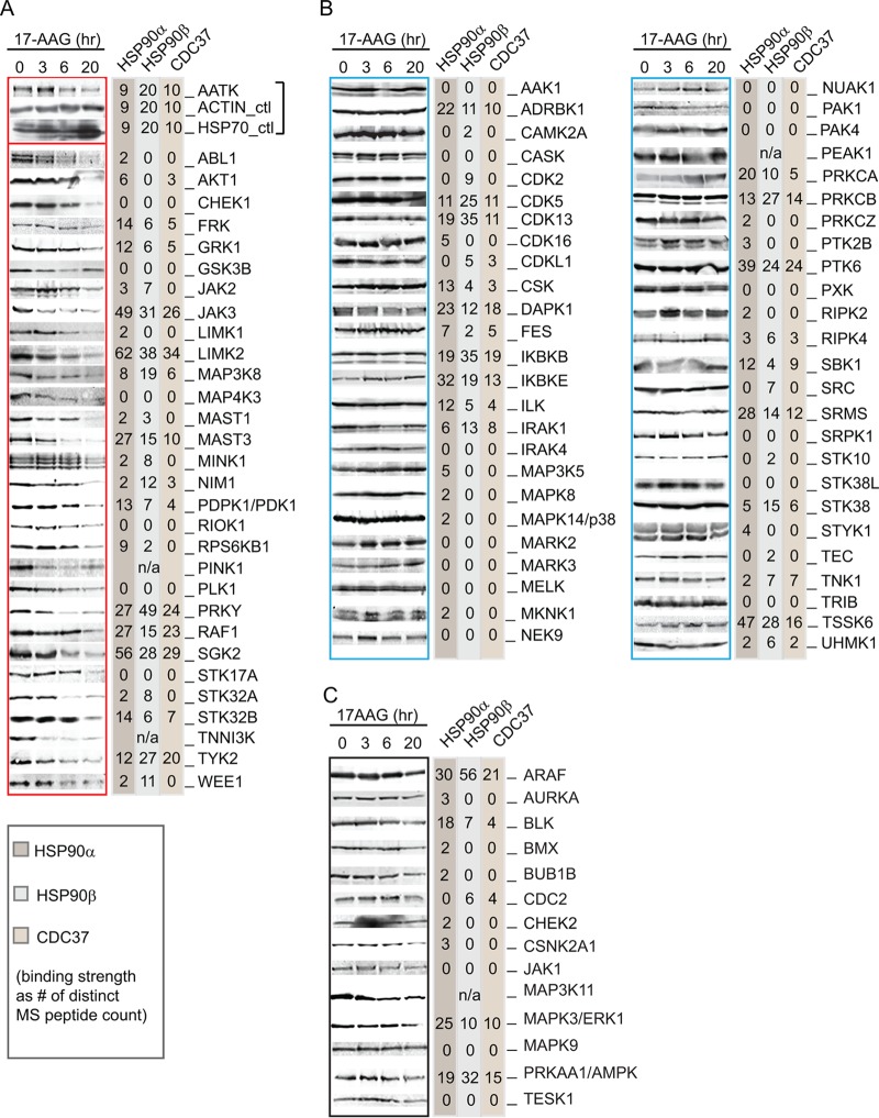
FIG 2.
HSP90 binding capacity and stability of protein kinases following 17-AAG treatment. Selected cytoplasmic protein kinases were individually tagged with an N-terminal FLAG and stably expressed in DLD-1 cells. Results from anti-FLAG Western blotting of cell lysate following a time course of 17-AAG inhibition are shown at the left of each panel (gel images were from a single experiment of cell treatment). Following anti-FLAG IP of the kinases from cell lysate, interacting proteins were identified by LC-MS/MS. The unique peptides identified by mass spectrometry for HSP90α, HSP90β, and CDC37 are shown at the right of each panel. For each membrane, anti-β-actin and anti-HSP70 blots were included as controls for sample loading and cellular response to inhibitor (representative control blots for AATK are shown). Kinases presented in panel A showed decreased expression upon 17-AAG treatment, while those listed in panel B remained at a constant expression level. (C) FLAG-tagged kinases whose stability after 17AAG treatment was inconclusive. These kinases are indicated as unclassified in Table S1 in the supplemental material.

