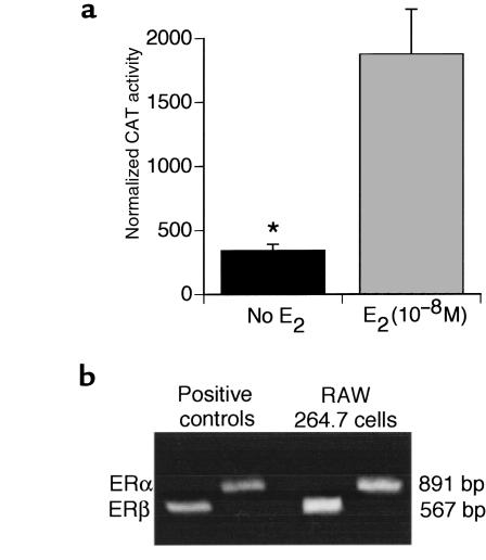
Figure 1.
RAW 264.7 cells express functional ERs and mRNA for ERα and ERβ. (a) Cells were transfected with a reporter plasmid that contained a CAT gene under the transcriptional control of an ERE driven by an SV-40 promoter, and were treated with either E2 or control vehicle. CAT activity was normalized to β-galactosidase activity to correct for variability in transfection efficiency and expressed as normalized CAT activity. Mean ± SEM of 3 experiments. *P < 0.05 vs. untreated cells. (b) RT-PCR amplification products of ERα and ERβ. Total RNA was extracted and reverse transcribed. The resulting cDNA was amplified using primers specific for ERα and ERβ. Two amplification products corresponding to ERα and ERβ were detected by ethidium bromide staining of agarose gels. Amplification of purified mRNA with ERα and ERβ primers in the absence of RT generated no detectable bands (data not shown).

