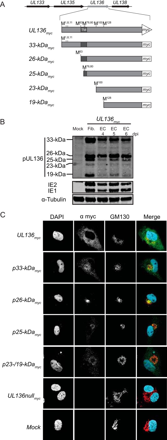
FIG 1 .

Expression and distinct localization of pUL136 in endothelial cells. (A) Schematic of the protein isoforms of UL136. (B) Expression of the pUL136 isoforms in ECs. HMVECs were either mock infected or infected with the UL136myc virus at an MOI of 2 and harvested at 4 to 6 dpi. As a positive control, MRC-5 fibroblasts were either mock infected or infected with the UL136myc virus at an MOI of 2 and harvested at 3 dpi. Lysates were immunoblotted with an antibody specific to the myc epitope tag. A monoclonal antibody to α-tubulin was used as a loading control. Fib., fibroblasts. EC, endothelial cells. (C) Distinct localization of the pUL136 isoforms in ECs. HMVECs were either mock infected or infected at an MOI of 3 with indicated viruses. At 5 days postinfection, the UL136 proteins were localized by indirect immunofluorescence using an antibody specific to the myc epitope tag or a Golgi apparatus marker, GM130. DAPI staining marks the nuclei. DAPI, blue. Myc, green. GM130, red.
