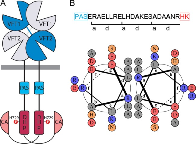
FIG 1 .
The PAS-HK linker of BvgS. (A) Schematic representation of the BvgS dimer. The receiver and HPt domains were omitted for clarity. The DHp domain, with the phosphorylatable His, and the catalytic and ATP-binding domains (CA) form the His kinase. (B) Amino acid sequence of the PAS-HK linker showing the a and d residues of the coiled coil, and representation by a helical wheel diagram using DrawCoil 1.0 (http://www.grigoryanlab.org/drawcoil/) as in reference 28. Hydrophobic residues are in grey, polar uncharged residues in orange, negative residues in red, and positive residues in blue. The abcdefg annotations correspond to the 3 heptads. PAS and HK represent the PAS and His-kinase domains, respectively.

