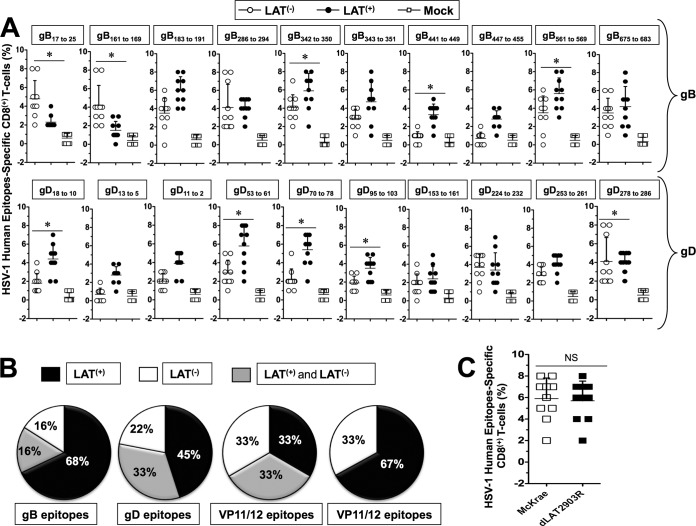
FIG 3.
Profiles of HSV-1 gB- and gD-derived human epitope-specific CD8+ T cells are detected in infected LAT+ TG versus LAT– TG. Three groups of HLA Tg rabbits Tg rabbits (n = 10) were ocularly infected with 2 × 10⁵ PFU/eye of the wild-type McKrae (LAT+), its LAT-null virus mutant dLAT2903 (LAT–), or the marker-rescued virus dLAT2903R counterpart (LAT+), as in Fig. 2. Both TG were harvested from each rabbit on day 35 (10 rabbits) postinfection. The 20 TG were pooled and treated with collagenase I, and the percentages of CD8+ T cells specific to HSV-1 human epitopes selected from glycoproteins B and D were determined in TG cell suspensions by FACS. (A) Percentages of HSV-1 human gD, gB, VP11/12, and VP13/14 epitope-specific CD8+ T cells detected during latency (day 35) in 10 LAT+ TG versus 10 LAT– TG infected HLA Tg rabbits. Each black and white circle represents individual HLA Tg rabbits. (B) Pie charts represent the frequencies of CD8+ T cells specific to HSV-1 human gD, gB, VP11/12, and VP13/14 epitopes detected in LAT+ TG compared LAT– TG. *, P < 0.05 (for the percentage of epitope-specific CD8+ T cells in LAT+ TG versus LAT– TG). (C) Percentages of HSV-1 gB342–350 epitope-specific CD8+ T cells detected in TG of rabbits infected with the marker-rescued virus dLAT2903R versus wild-type McKrae.

