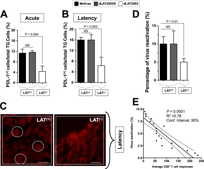
FIG 7.
HLA Tg rabbits infected with LAT+ virus have increased spontaneous virus shedding in tears and express high levels of PD-L1 molecules in TG compared to HLA Tg rabbits infected with LAT– virus. (A) HLA Tg rabbits infected with the wild-type McKrae (LAT+), its LAT-null virus mutant dLAT2903 (LAT–), or the marker-rescued virus dLAT2903R counterpart (LAT+). TG were removed 12 (Acute) or 35 (Latency) days after ocular infection. Half of the TG were treated with collagenase, and the other half were fixed, embedded in OCT, immunostained as indicated, and examined for expression of PD-L1 by confocal microscopy as described in Materials and Methods. Cell suspensions from LAT+ TG and LAT– TG harvested on five rabbits on day 12 (acute infection) or day 35 (latency) were stained with FITC-anti-human PD-L1 MAb (clone M1/42). The percentages of total TG-derived cells expressing PD-L1 detected by FACS in 10 LAT+ TG versus 10 LAT– TG during acute (A) and latent (B) phases are shown. (C) TG cells expressing significant detectable levels of PD-L1 were visualized by immunostaining in LAT+ TG versus LAT– TG during latency. (D) HLA Tg rabbits latently infected with LAT+ virus show increased spontaneous virus shedding in tears. HSV-1 shedding detected in tears was monitored for 30 days postinfection, as described in Materials and Methods. (E) Correlation between the percentage of positive tears and the average T cell responses during a 30-day monitoring period. *, P < 0.05 (ANOVA).

