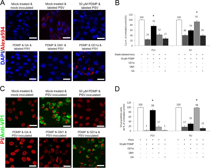
FIG 7.
PSV binding and infection are rescued by addition of GD1a. (A) The binding of AF-594-labeled PSV (MOI, 1,000 PFU/cell) was examined by confocal microscopy after pretreating LLC-PK cells with PDMP for ganglioside depletion followed by the addition of various free gangliosides. (B) Binding of [35S]methionine-cysteine-labeled PSV or RV (50,000 cpm) was measured by liquid scintillation counting after pretreating cells or not with PDMP for ganglioside depletion, followed by the addition of various free gangliosides. The levels of bound virus were expressed as a percentage of the value for the mock-treated, virus-inoculated control. (C) The effect of PDMP for ganglioside depletion followed by the addition of various free gangliosides on the infection of cells was assessed by immunofluorescence using monoclonal antibody specific for PSV capsid protein at 15 h postinfection. (D) The levels of PSV or RV antigen-positive cells expressed as a percentage of the values for the mock-treated, virus-inoculated control were quantified from three independent fields of view. All experiments were performed three independent times. Panels A and C show representative sets of results. The scale bars correspond to 20 μm. Error bars indicate SD from triplicate experiments. The asterisks in panels B and D indicate the P value (<0.05) obtained when comparing the PDMP treatment groups with PDMP treatment following ganglioside replenishment.

