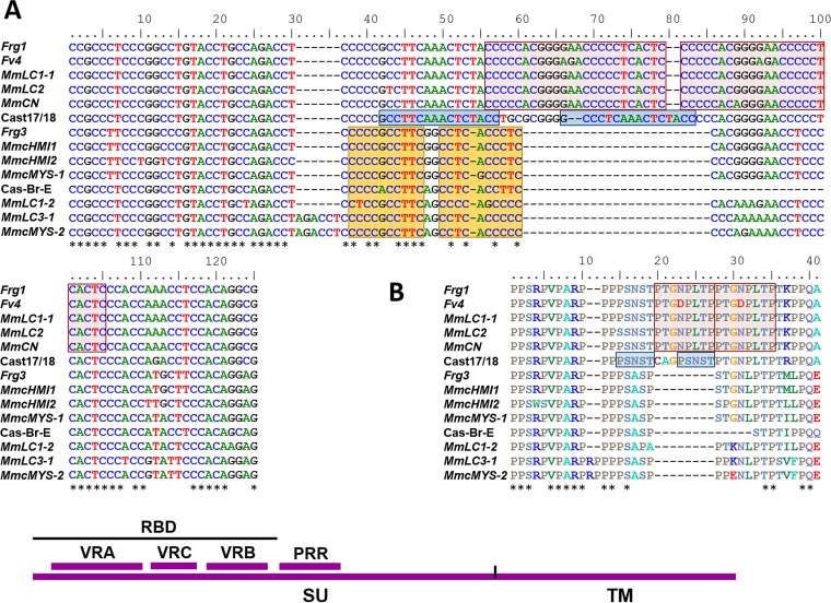
FIG 7.
Segmental duplications in the PRR regions of Cas subtype E-MLVs. Three different nucleotide duplications are marked by pink, blue, and yellow (A), and two amino acid duplications are marked by pink and blue (B). The nine Mm entries represent unique ERV PRR sequences amplified from M. m. castaneus or Lake Casitas (LC) mice. Asterisks mark sequence identities. At the bottom is a diagram of env showing the surface (SU) and TM domains, the RBD, the three variable domains within RBD (variable region A to variable region C [VRA-VRC]), and PRR.

