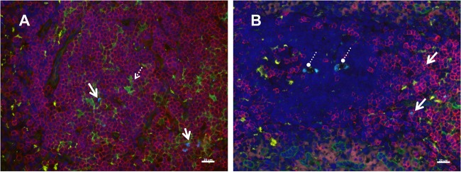
FIG 5.
Immunophenotype of SIV RNA+ cells 24 h after SIVmac251 inoculation. Cells containing vRNA were detected in tissue sections using in situ hybridization with fluorescently tagged antisense SIV-specific riboprobes and antibodies to phenotype cells using cell markers in tissues at 24 h p.i. (A) Inguinal lymph node; (B) spleen. Solid arrows, representative SIV RNA+ T cells (bright blue); these are often associated with p55+ (fascin-positive) DCs; dashed arrow in panel A, a vRNA+ T cell abutting a p55+ DC; circles with dashed lines in panel B, vRNA within macrophages. The pattern of vRNA in the macrophage cytoplasm (several discrete vRNA+ foci) is in a pattern consistent with the phagocytosis of vRNA+ T cells. Red, CD3+ T cells; green, p55+ endothelial cells and bone marrow-derived DCs; yellow, CD68+ macrophages; bright blue, SIV RNA; dark blue, DAPI staining of nuclear DNA. Bars = 20 μm.

