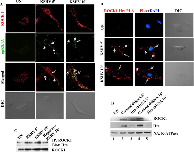
FIG 4.
Hrs facilitates recruitment of ROCK1 to the plasma membrane of infected cells. (A) Membrane localization of ROCK1 in KSHV-infected cells. HMVEC-d infected with KSHV for 5 and 10 min were incubated with anti-ROCK1 and anti-KSHV gpK8.1A antibodies. Localization of ROCK1 and gpK8.1A was analyzed by staining with Alexa 488- or Alexa 594-conjugated secondary antibodies. Arrows indicate gpK8.1A and ROCK1 localization at different times of infection. The differential interference contrast (DIC) image shown in the bottom panels corresponds to the fluorescence image. Magnification, ×80. (B) Colocalization of Hrs and ROCK1 visualized by in situ proximity ligation assay. Uninfected and KSHV-infected cells were subjected to PLA using a mouse anti-Hrs antibody and a rabbit anti-ROCK1 antibody. Nuclei were stained with DAPI. White arrows indicate fluorescent red dots representing the interaction between ROCK1 and Hrs. The corresponding DIC images are shown in the rightmost panels. Magnification, ×80. (C) Hrs interacts with ROCK1 in KSHV-infected cells. HMVEC-d were infected with KSHV for 5 and 10 min or with heparin-treated (100 μg/ml) KSHV for 10 min, and the cell lysates were immunoprecipitated with anti-ROCK1 antibody, followed by anti-Hrs or anti-ROCK1 immunoblotting. (D) Western blot showing the membrane association of ROCK1 in control and Hrs shRNA-transduced cells. Control and Hrs shRNA-transduced cells were infected with KSHV for 5 and 10 min, and the plasma membrane fractions of uninfected and infected cells were isolated and analyzed by Western blotting with antibodies for ROCK1, Hrs, and the plasma membrane marker Na, K-ATPase.

