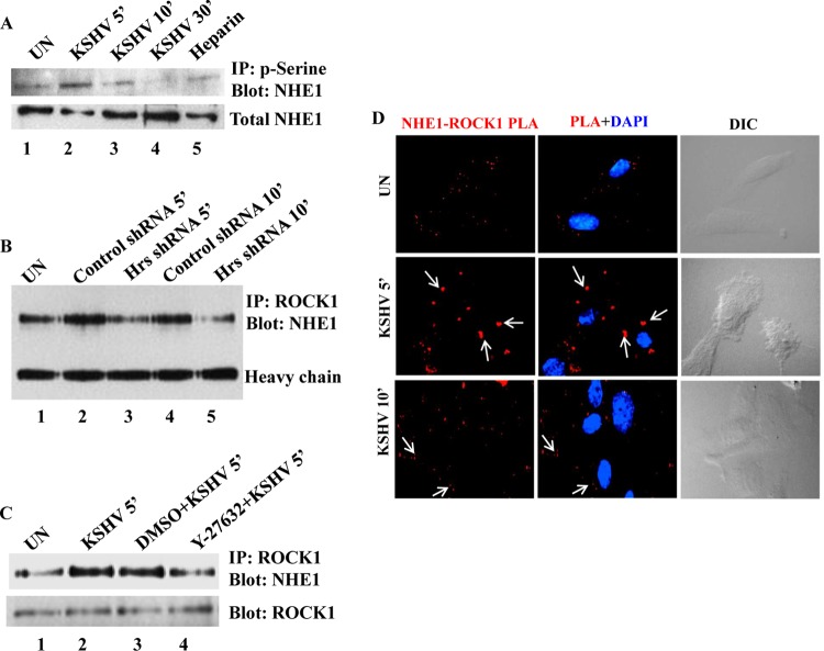
FIG 6.
ROCK1 interacts with and phosphorylates NHE1 in KSHV-infected cells. (A) Kinetics of NHE1 phosphorylation in KSHV-infected cells. HMVEC-d were left uninfected or infected with KSHV at an MOI of 20 for 5, 10, and 30 min or with heparin (100 μg/ml)-treated virus for 10 min, and the cell lysates were immunoprecipitated with antiphosphoserine antibody, followed by Western blotting with anti-NHE1 antibody. The bottom panel shows total cell lysates Western blotted with anti-NHE1 antibody. (B) Control and Hrs shRNA-transduced HMVEC-d were infected with KSHV for 5 and 10 min, and the uninfected cell lysates were immunoprecipitated using anti-ROCK1 antibody, followed by Western blotting with anti-NHE1 antibody. Heavy chain is shown to verify equal loading. (C) HMVEC-d left untreated or treated with vehicle DMSO or Y-27632 for 1 h were infected with KSHV for 5 min, and the cell lysates were immunoprecipitated with anti-ROCK1 antibody and Western blotted with anti-NHE1 antibody. The bottom panel shows the membrane stripped and reprobed with anti-ROCK1. (D) PLA showing the interaction between NHE1 and ROCK1 in the membrane of blebs. Uninfected and KSHV-infected HMVEC-d were fixed, permeabilized, blocked, and probed with antibodies to Hrs and ROCK1. Interaction of Hrs and ROCK1 in the infected cells was visualized by the red fluorescence dots (white arrows) using immunofluorescence microscopy. DAPI was used for nuclear staining. Magnification, ×80.

