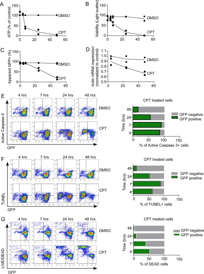
FIG 6.
Apoptotic cell death is associated with loss of protein and mRNA markers. Jurkat T cells stably expressing eGFP were treated with DMSO or CPT and assessed for cell viability by ATP content (A) and light scatter (B) over time. (C) Expression of eGFP was measured by flow cytometry. (D) Actin mRNA expression was assessed by qRT-PCR and expressed as mean CT values normalized to baseline DMSO control. (E to G) Cell death was assessed by active-caspase 3 expression (E), TUNEL (F), and Live/Dead viability stain (G) and compared between eGFP-positive and eGFP-negative cells. Values in panels A to D represent the mean (range) of two independent experiments. The results the panels E to G are representative of two independent experiments.

