FIG 2.
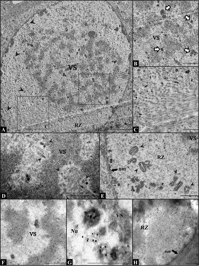
Electron micrographs of Sf9 cells transfected with vAc54KO or infected with wt AcMNPV. (A) Nucleus of a vAc54KO-transfected Sf9 cell at 24 h p.t. No capsid structures (with or without DNA content) exist in the VS. Black arrowheads indicate normal-appearing nucleocapsids in the RZ. (B) Partial view of the VS magnified from the cell shown in panel A, showing a large quantity of abnormal electron-dense bodies (indicated with white arrows) at the edge of the electron-dense mats. (C) Magnified view from panel A. Abnormally long, electron-lucent tubules can be found in the RZ. (D and E) Partial view of an Sf9 cell infected with wt AcMNPV at 24 h p.i. Nucleocapsids with DNA content are found in the VS (D), and ODVs are found in the RZ (E). Black arrowheads indicate normal nucleocapsids. (F to H) Immunoelectron microscopy analysis of vAc54KO-transfected Sf9 cells at 72 h p.t. (cells were labeled with BrdU at 12 h p.t.). Antiserum against BrdU (F), P6.9 (G), or VP39 (H) was used as the primary antibody. VS, virogenic stroma; RZ, ring zone; Nu, nucleus; nm, nuclear membrane. Scale bars, 2 μm (A) and 500 nm (B to H).

