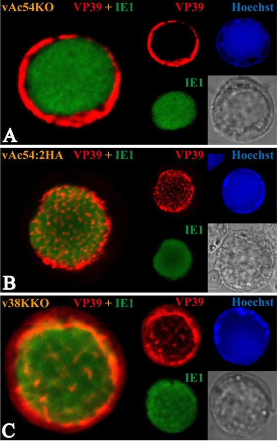
FIG 3.

Subcellular localization of the major capsid protein VP39. Sf9 cells were transfected with vAc54KO (A), vAc54:2HA (B), or v38KKO (C) bacmid DNA. Transfected cells were fixed at 36 h p.t. and incubated with antisera against VP39 (rabbit) and IE-1 (mouse) as the primary antibody mixture. The primary antibodies were visualized with Alexa Fluor 555-conjugated goat anti-rabbit (VP39; red) and Alexa Fluor 647-conjugated donkey anti-mouse (IE1; green) antibodies. Hoechst 33342 was used to visualize DNA-rich regions of the nucleus (blue).
