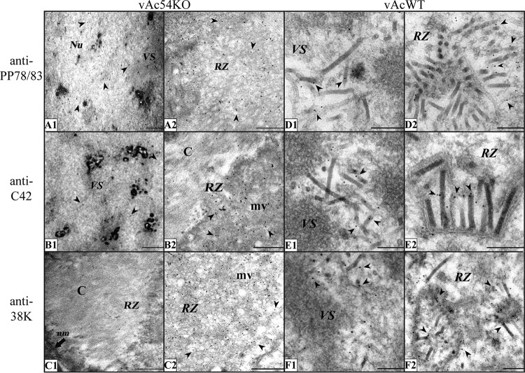
FIG 6.
Immunoelectron microscopy analysis of Sf9 cells transfected with vAc54KO or vAcWT bacmid DNA at 72 h p.t. using antisera against different nucleocapsid structural proteins. For the experiments shown in panels A to C, Sf9 cells were transfected with vAc54KO bacmid DNA. (A) Antiserum against PP78/83 was used as the primary antibody. The entire nucleus was equally stained with gold particles. (B) Antiserum against BV/ODV-C42 was used as the primary antibody. A minimum BV/ODV-C42 signal is visible in the VS; in the RZ, gold particles specifically colocalize with microvesicles but not with capsid structures. (C) Antiserum against 38K was used as the primary antibody. 38K mainly colocalizes with microvesicles; only a background level of gold particles was observed in the VS or on capsid structures. For the experiments shown in panels D to F, Sf9 cells were transfected with vAcWT. Signals for PP78/83 (D), BV/ODV-C42 (E), and 38K (F) specifically localize to the nucleocapsids. Arrowheads, gold particles; Nu, nucleus; nm, nuclear membrane; mv, microvesicles; C, capsid structures. Scale bars, 500 nm (A to C) and 200 nm (D to F).

