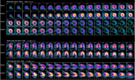
Fig. 1.

In July 2011, regadenoson rubidium82 rest–stress positron-emission tomographic images show a large, severe perfusion defect involving the mid-to-distal anterior, inferior, and apical left ventricular walls. The rest perfusion images (bottom row) notably show a pattern of enhanced radiotracer uptake in the mid-to-distal anterior and inferior walls and in the apex, similar to the spade shape on 2-dimensional echocardiograms and ventriculograms in patients who have apical-variant hypertrophic cardiomyopathy.
