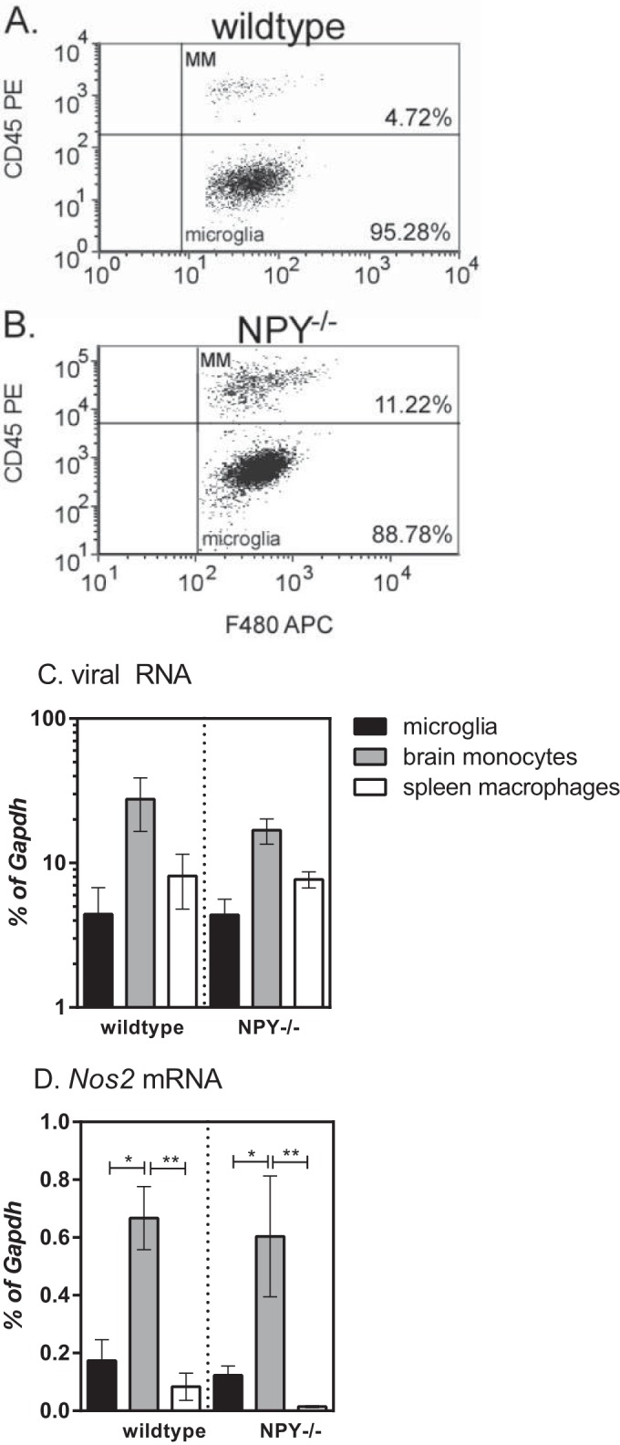
FIG 6.

Analysis of myeloid cells in the CNS following retrovirus infection in wild-type and NPY−/− mice. Brain tissue from mock- or Fr98-infected wild-type mice and Fr98-infected mice was removed at 21 dpi and homogenized into single-cell suspensions, and myeloid cells were isolated by Percoll gradient centrifugation. Individual cells were separated from the cell debris and doublets using forward and side scatter. Whole-cell populations were then gated for F4/80 expression. (A and B) F4/80-positive cells were analyzed for CD45 expression to identify brain monocytes (CD45hi) and microglia (CD45lo). Representative plots for Fr98-infected wild-type (A) and NPY−/− (B) mice are shown. (C and D) CD45lo F4/80+ microglia, CD45hi F4/80+ brain monocytes, and CD45hi F4/80+ spleen monocytes were isolated by sorting from the brains and spleens of Fr98-infected wild-type and NPY−/− mice at 3 weeks of age. RNA was isolated from each cell population and analyzed by real-time PCR for viral RNA (C) and Nos2 mRNA (D) expression. Gene expression was normalized to Gapdh mRNA expression for each sample. The data are from 8 samples per group, isolated in duplicate experiments. The data were analyzed by two-way ANOVA with Sidak's posttest. *, P < 0.05; **, P < 0.01. The error bars indicate SEM.
