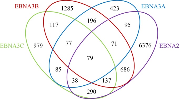
FIG 1.
Venn diagram showing colocalization of EBNA3A, EBNA3B, EBNA3C, and EBNA2 binding sites in LCLs. Sites on the human genome bound by EBV EBNA proteins were identified from ChIP-seq data as peaks relative to input using MOSAiCS. The numbers of bound sites and their extents of overlap are indicated for EBNA2, EBNA3A, EBNA3B, and EBNA3C.

