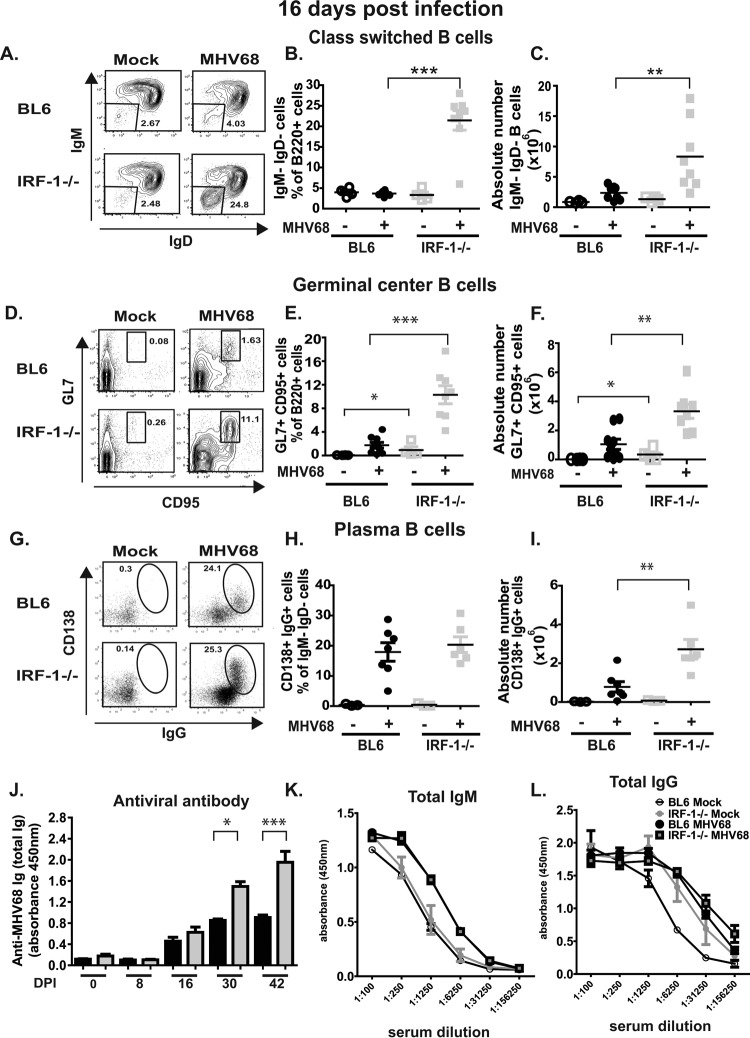
FIG 2.
IRF-1 restricts gammaherpesvirus-driven germinal center reaction during the establishment of viral latency. Mice of the indicated genotypes were infected as described in the Fig. 1 legend. (A to I) At 16 days postinfection, splenocytes harvested from individual animals were subjected to flow cytometry studies. Splenocytes were stained for surface expression of B220, IgM, and IgD (panel A shows representative results) to identify class-switched B cells (IgM negative [IgM−] IgD negative [IgD−]), which were quantified, and the result was expressed as a percentage of the number of B220+ cells (B) and as the absolute number per mouse spleen (C). Germinal center B cells (B220+) were identified by positive surface staining for GL7 and CD95 (panel D shows representative results), and the number of cells was expressed as a percentage of B220+ cells (E) and as the absolute number (F). Plasma cells were identified by surface CD138 and intracellular IgG staining (panel G shows representative results), and the number of cells was expressed as a percentage of IgM− IgD− B220+ cells (H) and as the absolute number (I). Each symbol represents the result for an individual spleen, and data from 2 to 4 independent experiments were pooled. (J) Sera collected from infected BL6 and IRF-1−/− mice at several times postinfection were diluted 1:50 and analyzed for virus-specific antibodies by ELISA. (K and L) Sera from mock- and MHV68-infected (16 days postinfection) mice were diluted, as indicated, and analyzed for total IgM (K) and IgG (L) levels. Data from two to four independent experiments were pooled.

