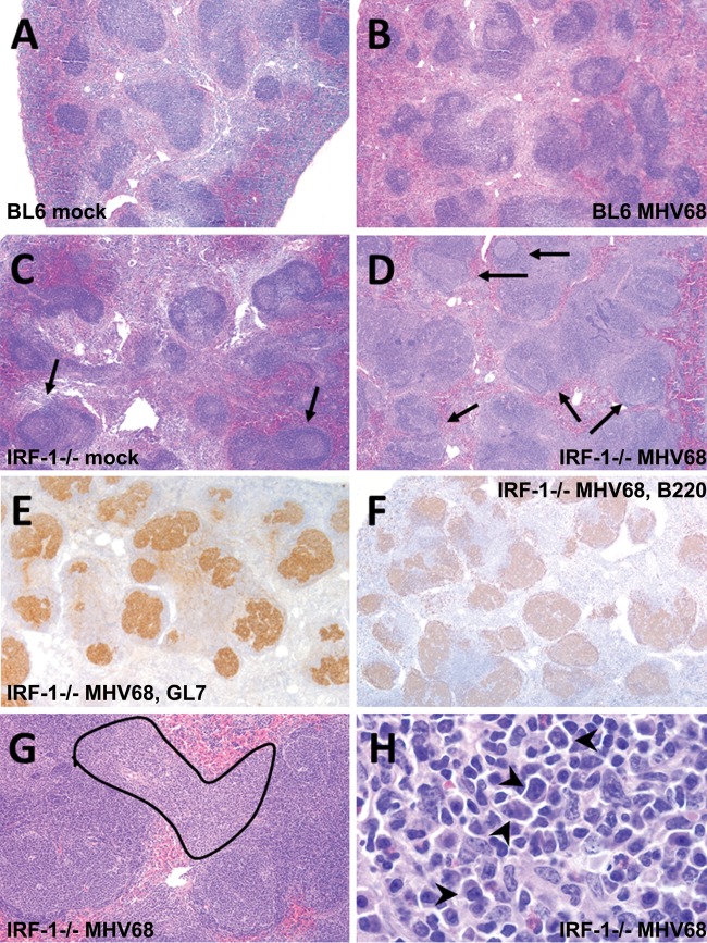
FIG 6.
Comparative spleen histology in BL6 and IRF-1-deficient mice. BL6 or IRF-1−/− mice were mock infected or infected with MHV68 as described in the Fig. 1 legend. Spleens were harvested at 42 days postinfection. (A and B) Mock-infected (A) and MVH68-infected (B) BL6 mice; (C and D) mock-infected (C) and MHV68-infected (D) IRF-1−/− mice; arrows, frequent germinal centers; (E and F) GL-7 (a germinal B cell marker) (E) and B220 (all B cells) (F) immunohistochemistry of the sample shown in panel D highlighting follicular hyperplasia; (G) PALS atypical lymphoid hyperplasia (outlined); (H) high-power magnification (500×) of the same lesion shown in panel G, showing atypical plasmacytoid cells (arrowheads). H&E staining was used. Magnifications, ×40 (A to D), ×100 (G), and ×500 (H).

