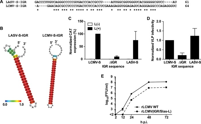
FIG 1.
Sequence plasticity associated with a functional S-IGR. (A, B) Comparison of the predicted structures of the S-IGRs of LASV and LCMV. The sequence alignment (A) and predicted structures (B) of the LCMV and LASV S-IGRs are shown. Predicted RNA secondary structures were determined using the CentroidFold web server (http://rtools.cbrc.jp). Each predicted base pair was colored using a heat color gradation from blue to red that corresponds to a base-pairing probability of from 0 to 1. Hyphens, gaps; asterisks, identical in LCMV and LASV S-IGRs. (C, D) The LASV S-IGR supports the production of LCMV infectious VLPs. The assay for the production of infectious VLPs was performed using the components of the LCMV S-MG, where the MG directed the expression of CAT and contained the S-IGR from either LCMV (LCMV-S) or LASV (LASV-S) or lacked an IGR sequence (ΔIGR). (C) Normalized CAT expression of transfected cells. The value for the WT MG was set to 100%. (D) Normalized VLP infectivity. The level of CAT expression of VLP-infected cells was divided by the level of CAT expression of transfected cells. The level of CAT expression in cells infected with VLPs produced by the WT MG was set to 1. Data represent the means ± SDs from three independent experiments. (E) Growth kinetics of rLCMV(IGR/Slas-L). BHK-21 cells were infected (MOI = 0.01) with either the rLCMV WT or rLCMV(IGR/Slas-L). Virus titers in the TCS were determined at the indicated times p.i. Data represent the means ± SDs from three independent experiments.

