FIG 4.
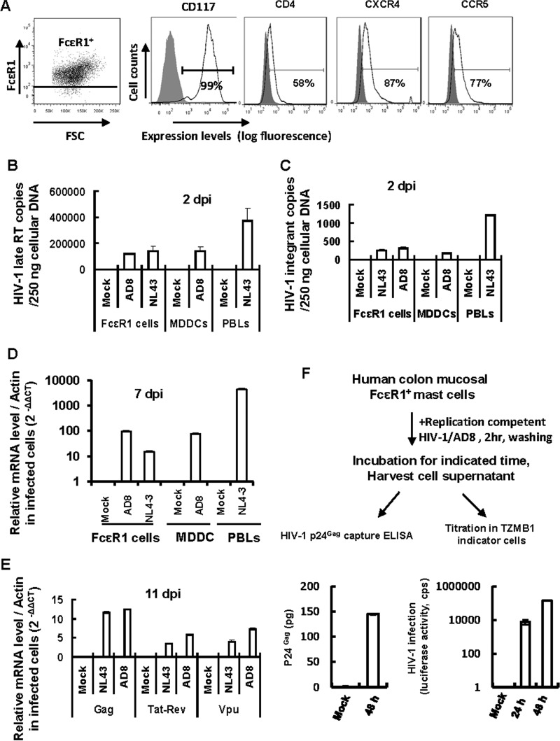
HIV-1 infection of gut mucosal mast cells. (A) Expression of HIV-1 (co)receptors as detected by immunostaining with specific antibodies and flow cytometry. (B to E) HIV-1 infection of mast cells was quantified by real-time PCR. Freshly isolated mucosal mast cells, autologous MDDCs, and PHA-P-activated PBLs were infected with wild-type HIV-1 AD8 or NL4-3 for 2 h, washed, cultured, and harvested at the indicated time. Mock infection was used as a control. Cellular DNA was extracted for quantification of late reverse transcriptase (late RT) products (B) and viral integrants (by Alu-PCR) (C), using 250 ng of cellular DNA for each sample, or total RNA was extracted after 7 or 11 days of infection, and the levels of HIV-1 total Gag RNA, multiply spliced RNA (Tat-Rev), and singly spliced RNA (Vpu) were quantified by real-time PCR and normalized to β-actin (D and E). (F) Mast cells are susceptible to productive HIV-1 infection. Purified FcεR1+ mast cells were infected with an amount of wild-type replication-competent HIV-1 AD8 equivalent to 5 ng p24gag for 2 h and then washed thoroughly. The cell culture supernatant was harvested at the indicated time for either HIV-1 p24gag capture ELISA to detect viral production or titration in TZMB1 indicator cells. Data are means and SD. Results are representative of four independent experiments.

