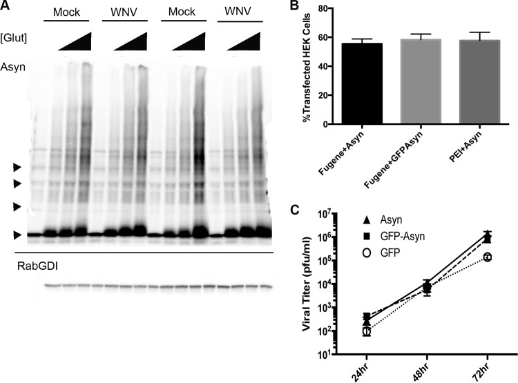
FIG 10.
WNV does not alter Asyn multimers in primary striatal neuron cultures. Primary striatal neurons were mock or WNV inoculated (MOI, 1) and treated with glutaraldehyde (Glut) at concentrations of 0%, 0.00125%, 0.0025%, or 0.005% upon harvesting at 24 h postinoculation. (A) Representative image of a Western blot obtained with a whole neuronal lysate labeled with antibody to Asyn. Each Western blot was also probed for Rab-GDI as a control for a cellular protein that does not cross-link. The image is representative of the images from three experimental replicates. Triangles on the left of the blot, expected molecular weights of the Asyn monomer, dimer, trimer, and tetramer (from bottom to top, respectively). (B) HEK293T cells were transfected with a plasmid expressing Asyn or green fluorescent protein (GFP)-Asyn. A mean ± SEM transfection efficiency of from 55 to 58% was calculated using IF analysis and antibody to Asyn or green fluorescent protein. Cells were analyzed at 24 h posttransfection, and means from 11 experimental replicates were calculated. PEI, polyethylenimine transfection reagent; Fugene, Fugene HD transfection reagent. (C) HEK293T cells were transfected with plasmids expressing green fluorescent protein alone, Asyn alone, or green fluorescent protein and Asyn. At 24 h posttransfection, cells were inoculated with WNV (MOI, 0.001) and the viral titer in the supernatants was determined at the indicated time points (n = 6 experimental replicates per treatment group).

