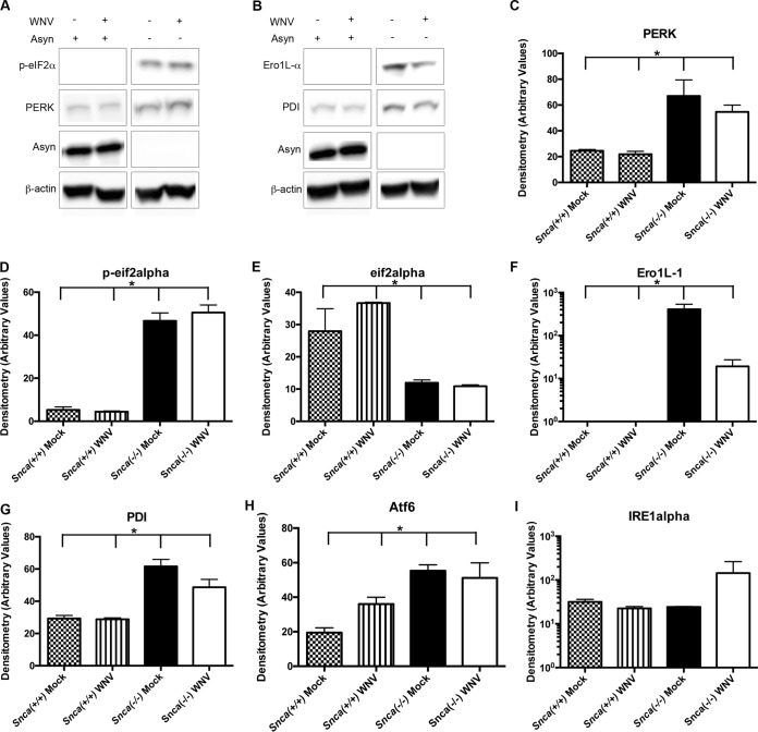
FIG 7.
Asyn expression modulates ER stress signaling. Primary cortical neurons derived from Snca+/+ and Snca−/− mice were mock inoculated or inoculated with WNV (MOI, 1) and harvested at 24 h for Western blot analysis of whole-cell lysates for ER stress signaling proteins. (A) Representative image of a Western blot probed with antibodies to phosphorylated eIF2alpha (p-eIF2alpha), PERK, Asyn, and β-actin. (B) Representative image of a Western blot probed with antibodies to Ero1L-1α, PDI, Asyn, and β-actin. Data are for three experimental replicates. (C to I) Semiquantitative densitometry analysis of mean band density corrected for by the density of β-actin for PERK (*, P = 0.006) (C), phospho-eIF2α (p-eIF2α; *, P = 0.011) (D), total eIF2α (*, P = 0.001) (E), Ero1L-1α (*, P = 0.0006) (F), PDI (*, P = 0.003) (G), Atf6 (*, P = 0.013) (H), and IRE1α (P = 0.2, no significant difference) (I). Data were obtained from three experimental replicates, and means were compared using a nonparametric ANOVA with the Kruskal-Wallis test.

