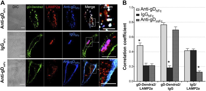
FIG 4.
Colocalization of gD and IgG with LAMP2A under ABB permissive and nonpermissive conditions. (A) Confocal slices of HeLa cells transiently expressing gp68 and gD-Dendra2 (green) and incubated for 2 h at 37°C under a 5% CO2 atmosphere with one of the three AF647-labeled IgGs (blue): anti-gDhFc (top), IgGhFc (middle), or anti-gDmFc (bottom). After fixation in 4% paraformaldehyde, cells were stained with AF568-labeled anti-LAMP2A (red). Regions of red (LAMP2A) and green (gD) colocalization appear yellow, regions of red (LAMP2A) and blue (IgG) colocalization appear purple, regions of green (gD) and blue (IgG) colocalization appear cyan, and regions of red (LAMP2A), green (gD), and blue (IgG) colocalization appear white. (B) Histograms comparing correlations at the 10-min (left) and 60-min (right) time points. Asterisks indicate a significant difference of colocalization compared to those of other members in the same category (P < 0.05).

