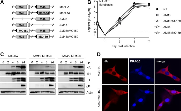
FIG 1.
Characterization of MCMV MC159 substitution mutants. (A) Schematic representation of the viruses used in this study. ◀, HA tag. (B) NIH 3T3 cells were infected with MCMV mutants at an MOI of 0.05 TCID50/cell. Supernatants were collected at the indicated days postinfection, and virus titers were determined. Means and standard errors of the means (±SEM) for experiments done in triplicate are shown. DL, detection limit. (C) NIH 3T3 cells were infected with MCMV M45HA, ΔM36::MC159, or ΔM45::MC159 at an MOI of 5 TCID50/cell. Cells were lysed at the indicated times postinfection, and viral protein expression was analyzed by immunoblotting. (D) NIH 3T3 cells were infected with an MOI of 2 TCID50/cell. Cells were fixed 6 hpi, and the subcellular localization of HA-tagged M45 and MC159 was analyzed by immunofluorescence. Nuclei were stained with DRAQ5.

