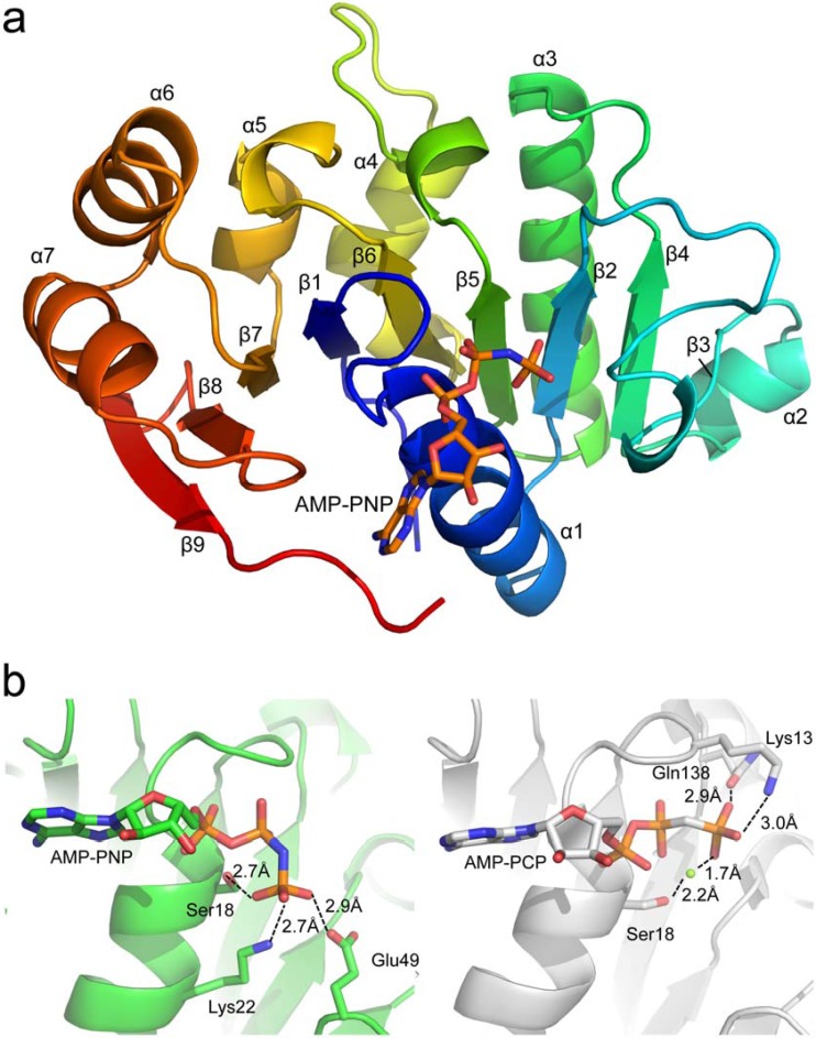
FIG 1.
(a) Global structure of STIV B204 shaded in rainbow colors from N terminus (blue) to C terminus (red). (b) Differences in orientation and stabilization of the γ-phosphate of the ATP analog (AMP-PNP or AMP-PCP) within crystal structures of STIV B204 (left; PDB code 4R2I) and STIV2 B204 (right; PDB code 4KFU), respectively. The magnesium ion is represented as a green sphere in STIV2 B204. The β-3 strand is represented by a β-bridge between the main-chain O atom of Tyr55 and the main-chain amino hydrogen atom of Val70.

