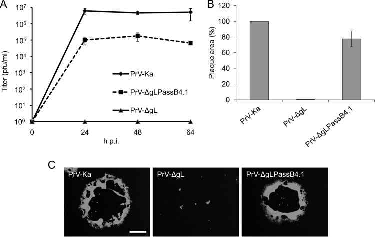
FIG 1.
In vitro growth properties of PrV-ΔgLPassB4.1. (A) For growth kinetics, RK13 cells were infected with PrV-Ka, PrV-ΔgL, or PrV-ΔgLPassB4.1 at an MOI of 0.5. Total cell lysates were harvested after 24, 48, and 64 h and titrated on RK13 cells. Given are mean values from three independent experiments with the corresponding standard deviations. (B) Cells were infected with PrV-Ka, PrV-ΔgL, or PrV-ΔgLPassB4.1 under plaque assay conditions and fixed at 2 days postinfection. Infected cells were visualized by indirect immunofluorescence with a monospecific anti-pUL19 serum, and plaque areas were measured. Plaque areas formed by PrV-Ka were set as 100%, and values for PrV-ΔgL and PrV-ΔgLPassB4.1 were calculated accordingly. Shown are the mean values and standard deviations from three independent experiments. (C) Representative images. Bar, 400 μm.

