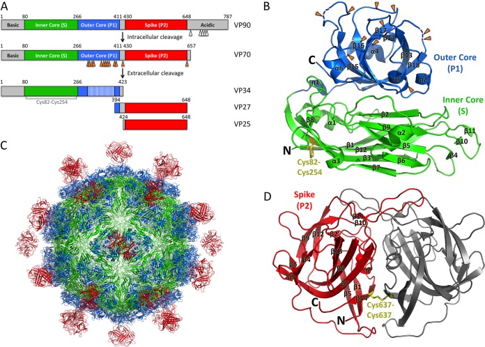
FIG 1.
Schematics and crystal structures of HAstV-1 CP core and spike. (A) Schematics of the HAstV-1 CP domain structure and proteolytic processing events. Caspase and trypsin cleavage sites are indicated with white and orange arrows, respectively. (B and D) Crystal structures of the HAstV-1 CP core (B) and spike (D). Trypsin cleavage sites are indicated with orange arrows. Disulfide bonds are labeled and colored yellow. N and C termini are labeled. (C) Model of mature T=3 HAstV-1 virion. Figures were made with PyMOL.

