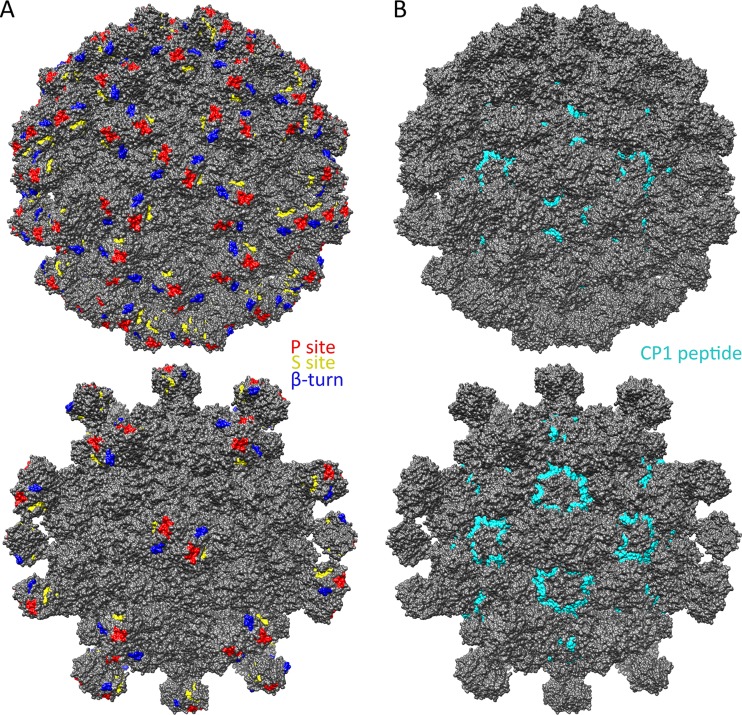
FIG 7.
Known and predicted functional sites on the surfaces of immature and mature T=3 HAstV-1 models. (A) Predicted receptor-binding sites, i.e., the P site (red), the S site (yellow), and the β-turn (blue), mapped onto immature (top) and mature (bottom) T=3 HAstV-1 models. (B) The CP1 peptide (HAstV-1 CP residues 80 to 138) (cyan) that binds complement C1q, mapped onto immature (top) and mature (bottom) T=3 HAstV-1 models. Figures were made with Chimera (40).

