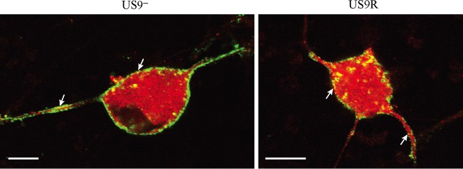
FIG 10.
Confocal micrographs showing colocalization of C capsids and envelope protein gE in dissociated rat neurons infected with US9− and US9R viruses at 24 hpi. The distributions of C capsids (red) and gE (green) in the cytoplasm of the cell body were similar between US9− and US9R viruses, showing no accumulation of capsids with envelope in the cytoplasm of the cell body, axon hillock, or proximal axons in the absence of pUS9. Bars, 10 μm.

