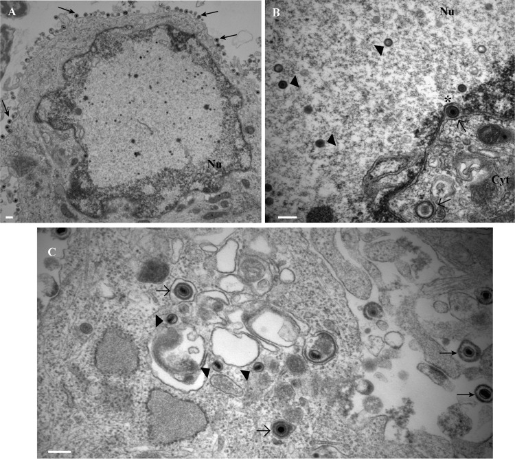
FIG 11.
Electron micrographs of rat neurons infected with HSV-1 US9− virus showing normal virus assembly and exit from the neuronal cell bodies in the absence of pUS9. (A) Lower magnification of neuronal cell body showing extracellular virions (arrows). (B) Capsids (arrowheads) in the nucleus and enveloped capsids (open arrows) in the space between inner and outer nuclear membranes (indicated with an asterisk) and in the cytoplasm close to the nucleus. (C) Enveloped (open arrows) and unenveloped (arrowheads) capsids in the cytoplasm of the cell body and extracellular virions (closed arrows). Bars, 200 nm.

