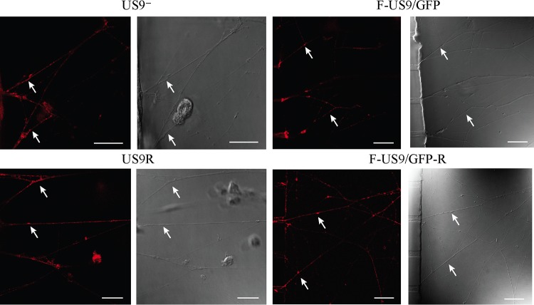
FIG 2.
Confocal micrographs of axons in the axonal compartment of neuronal cultures in microfluidic devices infected with US9 deletion (US9− and F-US9/GFP) and US9 repair (US9R and F-US9/GFP-R) viruses. Neonatal rat DRG neurons were dissociated, pelleted through a 35% Percoll gradient, and plated into the somal compartment of microfluidic devices. Cultures were incubated for 5 to 6 days to allow axons to grow into the axonal compartment. Vero cells were added to the axonal compartment 24 h prior to virus addition to the somal compartment. Foscarnet (100 μg/ml) was added to the axonal compartment 8 hpi to prevent secondary virus spread in Vero cells. The cultures were fixed at 22 hpi followed by immunostaining for C capsids (PTNC) and examined using a Leica SP5 confocal microscope. In the absence of pUS9, there was a reduction but not complete block in the anterograde axonal transport of capsids (arrows) to distal axons. Bars, 25 μm.

